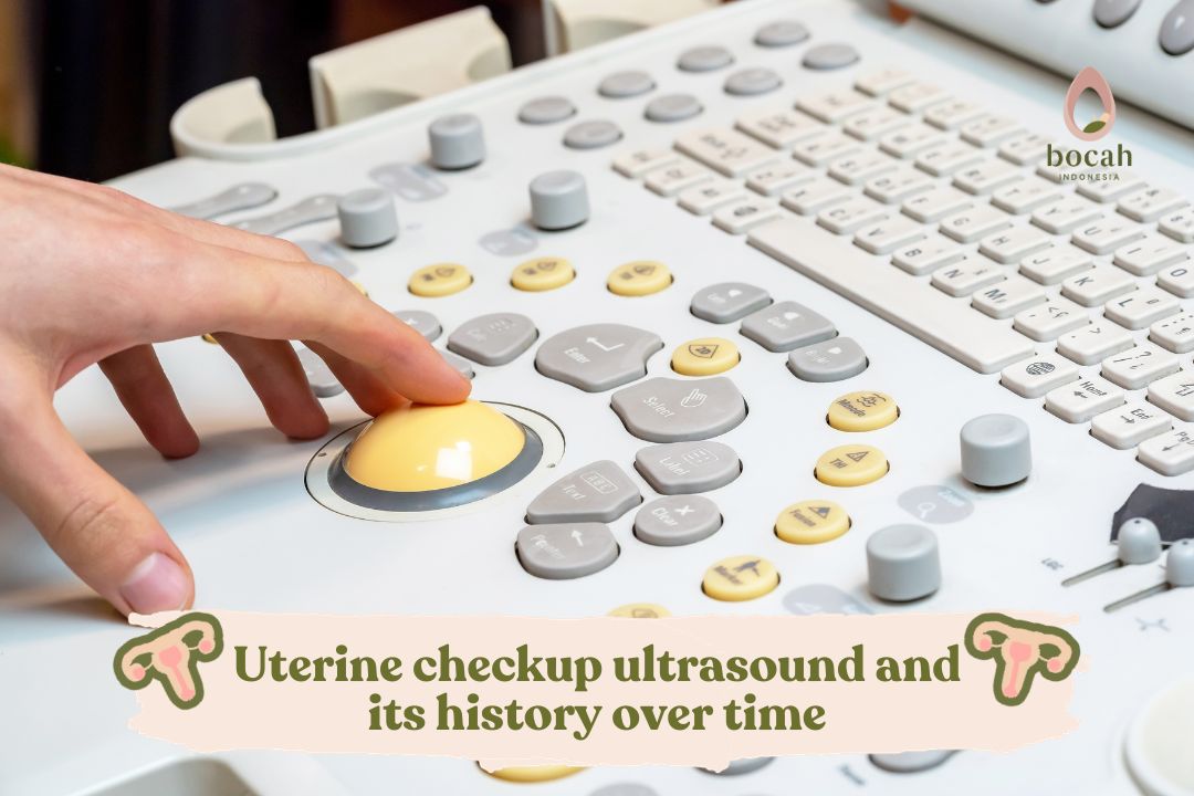
Pelvic ultrasound is performed to assess the condition of the uterus and other reproductive organs in women planning for pregnancy.
Ultrasound examination, also known as USG, is used to scan the body’s organs using high-frequency sound waves. However, this procedure does not produce radiation exposure, making it safe for all patients. There are several types of ultrasound examinations that can be performed.
One of the important examinations conducted to assess the condition of reproductive organs is ultrasound. Generally, ultrasounds used to check the uterus include transvaginal, transabdominal, and transrectal ultrasounds.
These three types of ultrasounds serve the same purpose, which is to detect the condition of the uterus, but the examination is conducted in different locations. Typically, transvaginal ultrasound is recommended for assessing the condition of the uterus.
But how did the history of uterine ultrasound examinations begin?
Mulai Promil Sekarang
The History of Ultrasound Over the Years
With the advancement of increasingly sophisticated technology, this development has also affected the field of healthcare, including ultrasound technology. In fact, the development of ultrasound has now reached 5D.
Here’s an explanation of the history of ultrasound development from its inception to the present day based on a journal published in Facts, Views, & Vision Obgyn.
In 1826, Jean Daniel Cooladon, a physicist from Switzerland, conducted research using church bells to communicate with his colleagues from one ship to another through water.
In 1880, Pierre Curie and Jacques Curie of France discovered the piezoelectric effect. They both agreed that ultrasound could produce images and be received in megahertz (MHz) frequencies.
Paul Langevin and Constantin Cholowsky of France developed a high-frequency echo sound device generated by ultrasound in 1915. This led to the creation of the hydrophone, which used transducers and quartz crystals previously discovered by Curie.
In 1943, Watson-Watt and his team used electromagnetic waves to create 2D ultrasound images. It was in 1949 that ultrasound was used to detect kidney stones and detect cancer in breast tissue.
Over time, Kratochwil used transvaginal ultrasound to detect fetal movement as early as 7 weeks of pregnancy. This occurred in 1967.
In the 1980s, ultrasound technology was introduced by Dr. Willyarto S. Wibisono, Sp.OG, who brought ultrasound to Indonesia from Germany.
Later, Kretztechnic developed transvaginal ultrasound for retrieving oocytes or eggs in IVF programs in 1985.
In the mid-1990s, advancing technology gave rise to the third-generation ultrasound, 3D and 4D, which played a crucial role in the fields of obstetrics and gynecology.
In 1995, Wilfred Feichtinger and Pieter Kemeter from Vienna explained that oocyte retrieval via transvaginal needle aspiration was now performed using a probe, which has become the standard technique in transvaginal ultrasound.
Finally, in the 2000s, the development of high-resolution transabdominal ultrasound machines and 3D and 4D transvaginal ultrasound continued. The use of ultrasound technology remains in practice today.
If you are advised to undergo a transvaginal ultrasound, there is no need to worry. This procedure typically takes approximately 5 minutes and does not cause discomfort. It also does not have any side effects on the body.
Do not hesitate to consult a doctor if you experience signs of infertility. Early examination helps you receive appropriate treatment and care to determine a pregnancy program. Let’s get examined with a transvaginal ultrasound at the nearest fertility clinic! It’s only a 5-minute* procedure, and it’s painless!
This article has been medically reviewed by Dr. Chitra Fatimah.
Source:
Drukker, L., et al. (2020). Introduction to artificial intelligence in ultrasound imaging in obstetrics and gynecology. Ultrasound Obstet Gynecol. 2020 Oct;56(4):498-505.
Campbell, S. (2013). A Short History of Sonography in Obstetrics and Gynaecology. Facts Views Vis Obgyn. 2013; 5(3): 213–229.
Dr. Shanty Olivia completed her medical school in 2005 and Obstetrics and Gynecology residency training in 2010 at the Faculty of Medicine, University of Indonesia. She joined a clinical fellowship program in Reproductive Endocrinology and Infertility at the same university and completed the program in 2018.
Latest posts by dr. Shanty Olivia F.J, Sp. OG, Subsp. FER (see all)