Spina bifida is a congenital birth defect that occurs when the spinal column and its nerves do not form completely. It is a type of neural tube defect (NTD) that commonly happens. In babies with spina bifida, a portion of the neural tube fails to develop fully.
What is spina bifida?
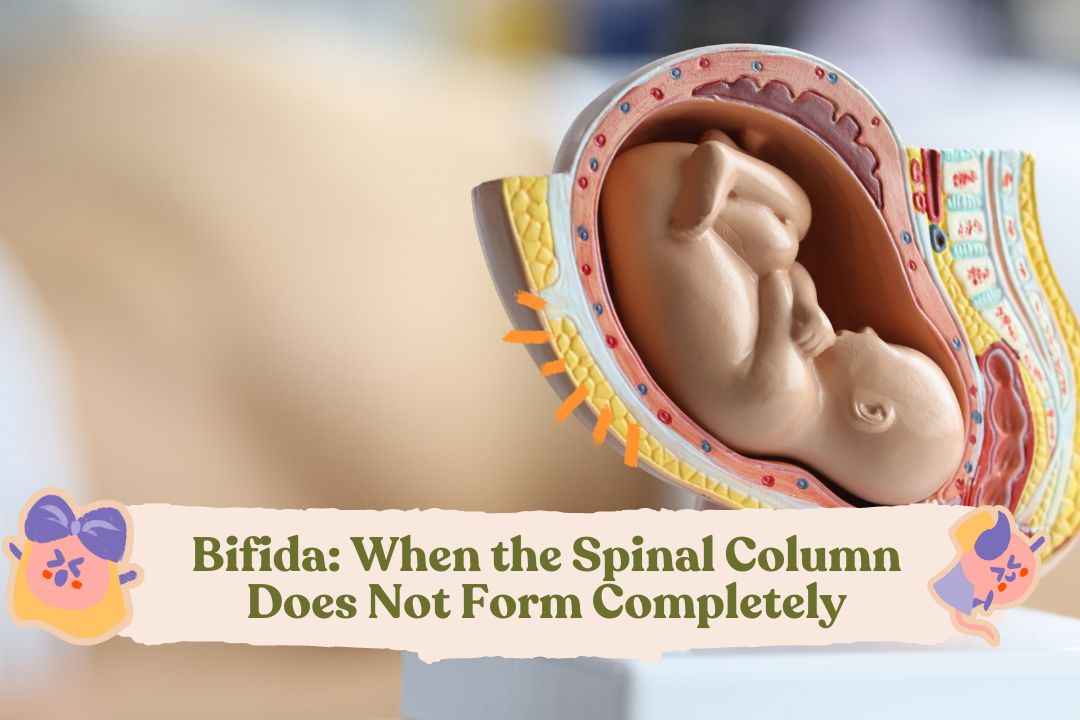
Spina bifida, which means “split spine” or “open spine,” is a congenital abnormality that occurs before birth. This condition happens when the neural tube, which eventually becomes the brain and spinal cord (a bundle of nerves along the spinal column), does not close completely. The neural tube starts as a small flat ribbon and develops into a tube. Normally, the neural tube closes during the first few weeks of pregnancy.
When the neural tube fails to close completely, the protective covering around the spinal cord, known as the spinal column, does not form properly, resulting in damage to the spinal cord containing the nerve fibers. Spina bifida can occur anywhere along the spinal column and can affect the development of the brain, brain membranes, spinal column, and its nerves. As a consequence, it can lead to motor, sensory, and other bodily function impairments.
Types of spina bifida
There are three main types of spina bifida known as follows:
1. Spina bifida occulta
Occulta means “hidden.” This type is also known as closed spina bifida because there are no visible openings on the back. The spinal cord and nerves are usually normal, with only minor abnormalities in the spinal bones.
Mulai Promil Sekarang
Some individuals may never experience any problems due to spina bifida occulta and may not even be aware that they have it. Others may show signs of abnormalities visible on the skin of the back, such as a small patch of hair or a dimple in the lower back, soft fatty lumps that can be felt, or port-wine nevi (reddish-purple spots).
2. Meningocele
This is a more severe type of spina bifida. The defect occurs when the outer part of the spinal column does not close completely, leaving a gap. The spinal cord is still in place, but its protective covering (meninges) may be damaged and pushed through the gap, forming a sac filled with cerebrospinal fluid. This sac is often covered by the skin.
In most cases, the spinal nerves are still normal or only slightly affected. This means that children with meningocele can have normal motor and sensory function in the lower limbs. Symptoms, if present, may include minor disruptions in bodily functions, such as back pain, leg weakness, and urinary or bowel dysfunction.
Myelomeningocele
This type, also referred to as open spina bifida, is the most common and severe form. When discussing spina bifida, it typically refers to myelomeningocele.
Similar to meningocele, the outer part of the spinal column does not close completely, leaving a gap. However, in this condition, both the meninges and the spinal cord protrude through the gap. Both structures bulge into a covered sac filled with cerebrospinal fluid, which has a thin membrane that can easily rupture, exposing its contents.
Because a portion of the spinal cord protrudes into the sac, it fails to develop properly, resulting in nerve damage. The resulting disabilities can range from moderate to severe. Most children with myelomeningocele will experience difficulty in movement and sensory impairments in the lower limbs. Paralysis and incontinence, which involves bedwetting or the inability to control bowel movements, are also common occurrences.
Causes and Risk Factors of Spina Bifida
The exact cause of spina bifida is not known, but it is believed to be related to genetic, environmental, and dietary factors that can affect the development of the fetal nervous system. One of the most extensively studied factors is the level of folic acid (vitamin B9) in pregnant women. Studies have found that a deficiency of folic acid during pregnancy increases the risk of spina bifida in the fetus.
Folic acid is thought to be involved in the process of forming fetal nerve cells, and therefore, it is recommended that pregnant women take daily folic acid supplements at least three months before conception and throughout pregnancy.
Other factors that can increase the risk of spina bifida include:
Ethnicity: Spina bifida is more commonly found in people of White and Hispanic ethnicity and is more prevalent in females than males.
Family history of neural tube defects: Couples who have previously had a child with a neural tube defect are at a higher risk of having another child with the same defect later on. The risk increases further if they have two children with the condition. Additionally, women born with spina bifida are also at an increased risk of giving birth to a baby with spina bifida. However, most babies with spina bifida are born to parents who do not have the condition.
Medications: Certain medications, such as the antiepileptic drug valproate, taken during pregnancy, can interfere with the body’s ability to absorb and process folic acid, leading to spina bifida.
Diabetes: Women with poorly managed diabetes are at an increased risk of having a baby with spina bifida.
Obesity: Being obese, especially before pregnancy, is associated with an increased risk of neural tube defects, including spina bifida.
Diagnosing Spina Bifida
Spina bifida can be diagnosed during pregnancy or after birth. During pregnancy, there are several tests to detect early abnormalities during routine prenatal visits. These include:
Alpha-fetoprotein (AFP) test: This simple blood test measures the amount of AFP produced by the fetus. Elevated AFP levels may indicate the presence of spina bifida in the baby.
Ultrasound (USG): Ultrasound can directly visualize fetal abnormalities, and spina bifida is often clearly visible through USG.
Amniocentesis: This test measures AFP levels in the amniotic fluid. Like the blood test, elevated AFP levels in the amniotic fluid may indicate spina bifida in the baby.
Sometimes, spina bifida may not be diagnosed until after the baby is born because the mother did not receive regular prenatal care or the ultrasound examination did not provide a clear image of the condition. After birth, doctors can diagnose newborns with spina bifida through visible abnormalities along the spinal column, such as a patch of hair on the skin or a dimple on the baby’s back. Doctors may also perform USG, X-rays, CT scans, or MRI to get a clearer picture of suspicious areas.
Health Issues and Complications Related to Spina Bifida
Each child with spina bifida may have a different level of disability. Some children may experience mild impairments, while others may have more severe disabilities. The severity of the impairments depends on:
The size and location of the spinal gap. Gaps in the upper spinal column are more likely to cause paralysis in the lower limbs and movement disorders compared to gaps in the middle or lower spinal column, which may only lead to incontinence.
The involvement of spinal nerves.
Common impairments and complications include:
Cognitive impairments: Spina bifida affects the developing spinal column and nerves, which can impact brain development. Areas of the brain involved in memory, learning, concentration, understanding, and language processing may be affected. Children may also experience difficulties in performing complex motor tasks such as tying shoelaces, which require visual and physical coordination. Statistically, 6 out of 10 children born with spina bifida have normal intelligence, although half of them may have specific learning disabilities.
Mobility impairments: The spinal cord allows information to travel up and down from the brain to the muscles and joints, controlling movement. Therefore, certain types of spina bifida can result in specific muscle paralysis, leading to limb developmental problems, especially in the legs. For example, clubfoot, where the baby’s foot is turned inward at the ankle, or scoliosis, where the spine curves abnormally in an “S” shape. The worst consequence of these mobility impairments is fragile bones or osteoporosis due to underuse of limbs.
Sensory impairments: The spinal cord and nerves also provide sensory information to the brain through touch. In certain types of spina bifida, nerve damage can lead to the loss of sensation in the pelvic and lower limb area. This can cause pressure sores and skin damage in babies who cannot feel the need to change their body positions.
Incontinence: Nerves that run through the spinal cord supply the bladder and bowel, ensuring that the muscles inside these organs can work to hold urine and feces inside the body and release them at appropriate times. In spina bifida, nerve disturbances cause children to experience urinary incontinence and bowel incontinence. When the bladder cannot empty completely (incomplete voiding), this condition puts the child at risk of urinary tract infections and kidney problems, such as infections or kidney stones.
In addition to the above impairments, there are several specific problems commonly found, such as:
Hydrocephalus or excess brain fluid: This condition can damage the brain and cause further issues. Many children with spina bifida and hydrocephalus have normal intelligence, but some may experience learning difficulties. In some babies, the bottom part of the brain is pushed downward towards the spinal cord. This condition, known as Arnold-Chiari malformation type 2, is associated with hydrocephalus. Hydrocephalus can cause additional symptoms soon after birth, such as irritability, seizures, sleepiness, pain, and difficulty feeding.
Allergy or sensitivity to latex: Babies with spina bifida are at an increased risk of developing allergies to latex-containing objects. Therefore, ensure that items used for the baby, such as gloves, catheters, and others, are latex-free.
Treatment for Spina Bifida
The treatment for spina bifida depends on the severity of the condition. Spina bifida occulta generally does not require any treatment, but other types of spina bifida may require surgery.
1. Prenatal Surgery
The function of nerves in babies with spina bifida can worsen after birth if left untreated. Prenatal surgery for spina bifida, also known as fetal surgery, is typically performed before the gestational age reaches 26 weeks. When done through open surgery, the doctor will open the mother’s uterus and repair the baby’s spinal cord. In certain cases, this procedure can be performed less invasively using a specialized instrument called a fetoscope, inserted into the uterus.
Studies have found that fetal surgery in children with spina bifida can reduce disability and decrease the need for crutches or other walking aids. Fetal surgery can also reduce the risk of hydrocephalus.
2. Postnatal Surgery
In meningomyelocele, surgery is often performed immediately after birth. However, the severity of disabilities after surgery depends on the presence of nerve tissue in the protruding sac. Subsequent treatment will be the same as for myelomeningocele.
Myelomeningocele requires surgery to close the gap in the baby’s back within 72 hours of birth. Performing surgery early can help minimize the risk of infection related to exposed nerves. It also helps protect the spinal cord from further trauma or injury.
During the procedure, a neurosurgeon will place the spinal cord and exposed tissues back into their proper position and then close them with muscle and skin. At the same time, the doctor may place a shunt in the baby’s brain to manage hydrocephalus. Additional surgeries may be needed to address other problems in the legs, pelvis, or spinal column.
Treatment for Complications
In addition to correcting anatomical abnormalities, children with spina bifida require care to optimize bodily functions, improve quality of life, and become more independent. Necessary treatments may include:
Mobility aids and walking aids, along with regular physiotherapy, will help children become more independent. Mobility aids can include crutches, walkers, or wheelchairs.
Routine evaluation of the bladder and bowel to reduce the risk of organ damage and diseases. Evaluation may involve X-rays, kidney examinations, ultrasound, blood tests, and bladder function studies. Specific treatments may include oral or suppository medications, enemas, catheter use to empty the bladder, surgery, or a combination of these.
Special equipment such as bath chairs, walking toilet seats, and standing aids can help with daily functions.
Despite the challenges, most spina bifida complications can be treated or at least managed to improve the quality of life for affected individuals.
Can Spina Bifida Be Prevented?
Yes, it can be prevented. Folic acid, in the form of supplements taken at least one month before pregnancy and continued during pregnancy (especially in the first trimester), significantly reduces the risk of spina bifida and other neural tube defects.
The current recommendations are that all women of reproductive age should consume 400 mcg of folic acid per day. Adult women planning to become pregnant or capable of becoming pregnant are advised to consume 400-800 mcg of folic acid per day. Women with a family history of spina bifida or neural tube defects should take 4 mg of folic acid per day.
In addition to taking supplements, women are also advised to consume foods rich in folate or fortified with folic acid. In food, folic acid is listed as folate, which is its natural form. However, the body cannot absorb folate as easily as synthetic folic acid. Additionally, most people do not get enough folate from their diets, making it necessary to take vitamin supplements to prevent spina bifida.
Some foods fortified with folic acid include bread, pasta, milk, and cereals. Natural food sources of folate include beans and legumes, citrus fruits, egg yolks, milk, avocados, and dark leafy greens, such as broccoli and spinach.
Conclusion
Spina bifida is associated with several complications that affect the quality of life for the child and residual symptoms involving various organ systems. Therefore, couples planning pregnancy need to understand this condition and its long-term impacts and take concrete steps to reduce the risk of its occurrence from the beginning.
Ask Mincah
[fluentform id=”31″]
Source:
Brea CM, Munakomi S. Spina Bifida. [Updated 2023 Feb 12]. In: StatPearls [Internet]. Treasure Island (FL): StatPearls Publishing; 2023 Jan-. URL: https://www.ncbi.nlm.nih.gov/books/NBK559265/
March of Dimes. [Last reviewed Jul 2021]. Spina bifida. URL: Spina bifida | March of Dimes.
Mayo Clinic. [Last reviewed 08 Jan 2022]. Spina bifida. URL: https://www.mayoclinic.org/diseases-conditions/spina-bifida/symptoms-causes/syc-20377860#:~:text=Overview,the%20tissues%20that%20enclose%20them.
Opšenák R, Richterová R, Kolarovszki B. Etiology and Pathophysiology of the Spina Bifida [Internet]. Spina Bifida and Craniosynostosis – New Perspectives and Clinical Applications. IntechOpen; 2021. Available from: http://dx.doi.org/10.5772/intechopen.97467
After graduating from the Faculty of Medicine, University of Indonesia, dr. Fiona served as a Non-Permanent Employee (PTT) doctor from the Indonesian Ministry of Health in a remote village in Luwuk-Banggai Regency, Central Sulawesi. This experience led her to continue her master's degree in International Health at Gadjah Mada University, Yogyakarta.