Let’s Understand the Differences Between Transvaginal Ultrasound and HSG Early On
Knowing the difference between Transvaginal Ultrasound (USG) and Hysterosalpingography (HSG) for a pregnancy program is crucial. Find out the distinctions here.
Both transvaginal ultrasound and HSG can be used to evaluate the reproductive health of women, involving an assessment of the structures from the vagina, cervix, uterus, fallopian tubes, to the ovaries.
To better understand the differences between these two types of examinations and how they can support the Mother’s pregnancy program, read more in this article.
Understanding Transvaginal Ultrasound for Conception
Transvaginal ultrasound (transvaginal ultrasonography) is a medical examination commonly performed on women undergoing a pregnancy program or facing difficulty conceiving.
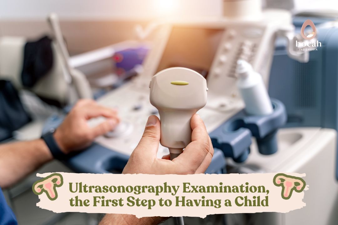
This examination involves the use of a special probe inserted into the vagina to obtain a closer and more detailed view of the female reproductive organs.
Tanya Mincah tentang Promil?
Objectives of Transvaginal Ultrasound Examination
1. Detecting ovulation
Transvaginal Ultrasound helps determine when ovulation occurs by monitoring the development of ovarian follicles. This can assist couples trying to conceive in identifying the right time for sexual intercourse.
2. Evaluation of reproductive organ structures
Examining the size, shape, and position of the uterus to ensure conditions that support pregnancy.
Assessing the number, size, and condition of the ovaries to ensure the health and availability of eggs.
3. Fetal health monitoring
If pregnancy has occurred, transvaginal ultrasound can be used to monitor fetal growth and development, as well as detect potential issues such as structural abnormalities or abnormal growth.
Transvaginal Ultrasound Examination Process
Before starting the examination, the doctor will provide guidance on actions that need to be taken by the mother during the ultrasound procedure. If necessary, the mother will be asked to empty or fill the bladder as needed to facilitate the examination.
If a full bladder is required, it is recommended to increase water intake starting one hour before the examination. .
Then, a lubricated probe is inserted into the vagina. This probe can provide a more accurate image due to its closer proximity to the reproductive organs.
After that, ultrasonic waves are emitted through the probe to the reproductive organs, and the reflection of these waves is used to create a detailed image of the uterus, ovaries, and other structures.
Information Obtained from Transvaginal Ultrasound
Transvaginal Ultrasound can confirm the presence of pregnancy and determine the gestational age more accurately. Then, this examination can also assess the condition of the uterus, ovaries, and fallopian tubes to identify potential issues or abnormalities.
In addition to conception, transvaginal ultrasound is often performed during pregnancy to examine the fetus and can also help determine the gender of the fetus.
It is important to note that transvaginal ultrasound is an examination generally safe to perform. The information obtained from this examination can help doctors and parents better plan and manage pregnancy more effectively.
Understanding Hysterosalpingography (HSG) for Conception
Similar to transvaginal ultrasound, Hysterosalpingography (HSG) is also performed for mothers undergoing a pregnancy program (promil) or facing difficulty conceiving. The main purpose of HSG is to evaluate the health of the uterus and fallopian tubes.
Objectives of HSG Examination for Conception
1. Evaluating the Uterus
HSG is used to assess the structure and condition of the uterus. This includes examining the shape, size, and condition of the uterine walls and detecting abnormalities such as polyps or fibroids.
2. Examining the Fallopian Tubes
This examination allows doctors to evaluate whether the fallopian tubes are open or experiencing blockages. The openness of the fallopian tubes is crucial to ensure the passage of sperm and eggs, allowing fertilization.
3. Detection of Structural Abnormalities
HSG helps detect structural abnormalities in the uterus or fallopian tubes that may affect the couple’s ability to conceive.
HSG Examination Process for Conception
During HSG, the doctor inserts a catheter into the uterus through the cervix. Contrast material visible on X-rays is then injected into the uterus, and radiographic images are taken.
The doctor will also use X-rays to observe the movement of the contrast material in the uterus and fallopian tubes. This allows the evaluation of the smooth passage of sperm and eggs.
Information Obtained from HSG for Conception
HSG provides information about whether the mother’s fallopian tubes are open or experiencing blockages. Open tubes facilitate the journey of sperm and eggs to meet.
HSG can also help doctors identify abnormalities such as polyps, fibroids, or a uterine septum that may affect the uterus’s ability to support pregnancy.
HSG examination can help determine the cause of infertility in couples experiencing difficulty getting pregnant.
HSG is an essential diagnostic tool in evaluating fertility and helps plan appropriate treatment strategies.
Although mothers may experience discomfort during the examination, HSG is generally considered a relatively safe and effective procedure in providing crucial information related to women’s reproductive health.
Differences Between Transvaginal Ultrasound and HSG
The fundamental difference between transvaginal ultrasound and HSG lies in the examination method. The examination transvaginal ultrasound uses ultrasound waves transmitted through the vagina to create images of women’s reproductive organs.
Meanwhile, HSG uses X-rays and contrast material to produce clearer images of the uterus and fallopian tubes. Sometimes, both of these examinations are performed simultaneously.
When transvaginal ultrasound indicates suspicion of fallopian tube blockage or abnormalities in the shape of the uterus, the doctor may decide for the mother to proceed with an HSG examination to obtain a clearer and more detailed picture.
To determine which examination is needed, it should be consulted with the doctor. The doctor will consider other examination results, the symptoms experienced by the mother, and overall health information.
Therefore, consult directly with a doctor to get a more detailed explanation and recommendations that are suitable for the mother’s health condition.
Aspiring for the presence of a child and wanting to try a pregnancy program, Dads and Moms can visit the Bocah Indonesia website.
This article has been medically reviewed by Dr. Chitra Fatimah.
Source:
- Toufig, H., et al. (2020). Evaluation of Hysterosalpingographic Findings Among Patients Presenting with Infertility. Saudi Journal of Biological Sciences, 27(11), pp. 2876–2882. https://pubmed.ncbi.nlm.nih.gov/33100842/
- Waheed, K., et al. (2019). Hysterosalpingographic Findings in Primary and Secondary Infertility Patients. Saudi Medical Journal, 40(10), pp. 1067–1071. https://www.ncbi.nlm.nih.gov/pmc/articles/PMC6887874/
- Panzone, J., et al. (2022). Transrectal Ultrasound in Prostate Cancer: Current Utilization, Integration with mpMRI, HIFU and Other Emerging Applications. Cancer Management and Research, 14, pp. 1209–1228. https://pubmed.ncbi.nlm.nih.gov/35345605/
- Drukker, L., Noble, J., & Papageorghiou, A. (2020). Introduction to Artificial Intelligence in Ultrasound Imaging in Obstetrics and Gynecology. Ultrasound in Obstetrics & Gynecology, 56(4), 498-505. https://pubmed.ncbi.nlm.nih.gov/32530098/


