With a 4D ultrasound examination, Moms and Dads will see the baby-to-be very clearly. Find out how to read the results of a 4D ultrasound here!
The need for a 4D ultrasound depends on Moms and Dads’ desire. 4D ultrasounds are generally not part of routine examinations with obstetricians, but they are often requested by couples who want a clearer picture of the baby’s condition in the womb.
This type of ultrasound provides Moms and Dads the opportunity to see the baby’s face and movements in a more real-time and clear manner. Find out more here to better understand the function and how to read the results of a 4D ultrasound.
What Is 4D Ultrasound (Ultrasonography)?
Ultrasound is a pregnancy examination that uses high-frequency sound waves to create images of the inside of the body. This technology first appeared in the form of 2D scans in the late 1950s but became more widely used for monitoring fetal development since the late 1970s.
With the advancement of technology, ultrasound examinations have become more sophisticated, introducing 3D and 4D (three-dimensional and four-dimensional) scans.
Mulai Promil Sekarang
With 3D ultrasound, doctors can see the fetus more clearly, and with 4D scans, this technology allows doctors to see finer details, such as the baby’s facial features and movements.
4D ultrasounds even enable doctors to assess how the central nervous system is developing and can help detect issues like cleft lip. All of this aids doctors in providing the best care for Moms and their babies during pregnancy.
Information Obtained from 4D Ultrasound
Facial features and expressions
In 4D scans, Moms can see the baby’s face more clearly, including expressions and subtle movements. This allows Moms to feel closer to the baby and see how the baby might look after birth.
Fetal movements
4D ultrasounds provide a fourth-dimensional aspect, allowing Moms and Dads to see fetal movements in real-time. Watching the baby move can be a joyful experience and provides insight into fetal activities in the womb.
Another advantage is that 4D ultrasounds can record the baby’s movements, such as swallowing, grimacing, and blinking, providing additional information about fetal development.
Gender determination
With clearer images, doctors may be able to determine the baby’s gender. However, this still depends on the baby’s position during the scan.
Heartbeat
Although the primary focus of 4D ultrasounds is visual, some equipment can also provide information about the fetal heartbeat. A healthy heartbeat is a positive sign of fetal development.
Organ structures
4D ultrasounds can help detect the structures of the baby’s organs, such as the face, hands, feet, and other organs. This can provide a more detailed picture of fetal physical development.
4D Ultrasound Includes 3D Ultrasound
4D ultrasounds include 3D (three-dimensional) images that display the baby from various angles and 4D (four-dimensional) images that add the dimension of time to observe the baby’s movements directly.
Understanding Terminology in Reading 4D Ultrasound Results
There are some terms that you may hear when undergoing a 4D ultrasound examination. So that you won’t be confused by the terms mentioned by the doctor, here are some terms in 4D ultrasound examinations and their meanings:
Gestational Age (GA) or pregnancy age
This refers to the estimated age of the fetus when the ultrasound is performed. Doctors use measurements of the fetus’s head diameter, leg length, and arm length to obtain a more accurate estimate of age.
Gestational Sac (GS) or pregnancy sac
The pregnancy sac appears in the first trimester and displays a round, black sac. This provides an initial visual representation of the success of fetal development.
Biparietal Diameter (BPD) or skull diameter
This term refers to the measurement of the fetus’s skull bone. BPD examination is usually done in the second and third trimesters to get a more detailed picture of the baby’s physical development.
Head Circumference (HC) or head circumference
The fetal head circumference provides information about the size of the baby’s head. This measurement is important as a healthy and proportionate head is a vital indicator of normal fetal growth.
Crown-Rump Length (CRL) or crown-rump length
CRL measures the length of the fetus from the top of the head to the bottom. This is one of the first parameters measured and helps confirm the exact gestational age.
Abdominal Circumference (AC) or abdominal circumference
The fetal abdominal circumference provides an estimate of the baby’s belly size. This measurement helps doctors evaluate overall fetal growth and development.
Femur Length (FL) or femur length
Femur length measures the length of the fetal thigh bone. This provides an overview of bone development and the baby’s leg structure.
Fetal Heart Rate (FHR) or fetal heart rate
FHR reflects the baby’s heart rate in the womb. This is an important parameter for assessing the fetal heart’s health.
Estimated Due Date (EDD) or estimated due date
EDD provides an estimate of when you may give birth. It is calculated based on information from your menstrual date and helps plan for childbirth preparations.
Last Menstrual Period (LMP) or last menstrual period
LMP is used to determine the gestational age by counting from the first day of your last menstrual period. This provides a common basis for pregnancy monitoring.
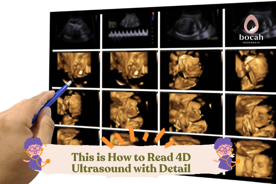
How to Read 4D Ultrasound Results
In a 4D ultrasound examination, three colors typically appear: gray, black, and white. Each color has a different meaning, here’s an explanation:
White Color: indicates hard body parts like bones. If you see white, it could mean the ultrasound is showing the baby’s bones.
Black Color: represents fluid, especially amniotic fluid around the baby. If the ultrasound shows black areas, it could indicate where the amniotic fluid is located.
Gray Color: indicates various soft tissues of the baby’s body. For example, gray color can represent how the baby’s body structure looks.
These color differences occur because the ultrasound sends sound waves and reflects them back. White color appears because sound waves bounce back more from hard surfaces, like bones.
However, it’s important to note that ultrasound cannot see parts of the body filled with air, such as the lungs, well. This is because sound waves do not work well when passing through air. That’s how you read 4D ultrasound results.
We hope this information helps you understand 4D ultrasound examinations. Information about fertility programs and IVF programs can be found on Bocah Indonesia.
This article has been medically reviewed by Dr. Chitra Fatimah.
source:
Benson, C.B. & Doubilet, P.M. (2018). Fetal Measurements: Normal And Abnormal Fetal Growth And Assessment of Fetal Well-Being. Diagnostic Ultrasound, 42, pp. 1443–1464. https://www.ncbi.nlm.nih.gov/pmc/articles/PMC2878887/
Grantz, et al. (2018). Fetal Growth Velocity: The NICHD Fetal Growth Studies. American Journal of Obstetrics and Gynecology, 219(3), e1–285.e36. https://pubmed.ncbi.nlm.nih.gov/29803819/
E.I. Medical Imaging. (2018). Ultrasound Basics: How to Read an Ultrasound Image.
Very Well Family. (2020). Early Pregnancy Ultrasound Results.