Follicle Monitoring in IVF Programs
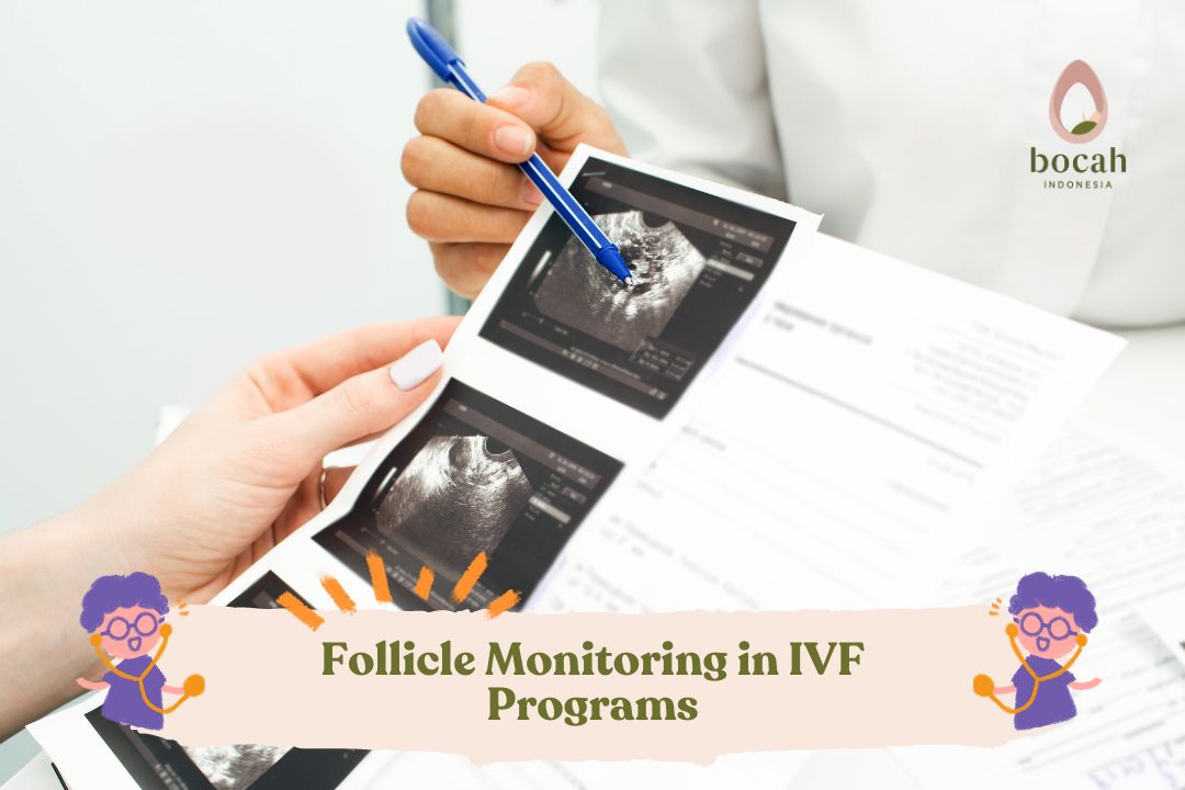
Monitoring the size of follicles during ovarian stimulation is very important to determine the woman’s response to the treatment and to optimize the number of mature eggs and the timing of their retrieval.
In the field of assisted reproductive technology (ART), understanding the significance of the size of egg follicles plays a crucial role in the success of pregnancy. For individuals or couples starting an in vitro fertilization program, understanding the development and size of ovarian follicles is crucial to unravel the complexity of the process.
What is a follicle in an IVF program?
A follicle is a small fluid-filled sac located within a woman’s ovary. Each follicle contains an immature egg cell, also known as an oocyte. In a natural menstrual cycle, several follicles begin to develop, but usually only one will mature and release an egg during ovulation. However, in an IVF program, the goal is to stimulate the ovaries to produce multiple mature follicles, each containing an egg ready for fertilization. The retrieval of these eggs should not be too early (when the eggs are not yet mature) or too late (when the eggs are mature and have started to degenerate).
This can be achieved by administering fertility medications through the process of ovarian stimulation. It is important to minimize the risk of Ovarian Hyperstimulation Syndrome (OHSS) during this process. This condition can be potentially dangerous if the ovarian response to stimulation is excessive, indicated by the development of more than 20 follicles and/or high estrogen levels in the blood before the egg retrieval procedure.
Why is it important to monitor follicle development?
Monitoring follicle development during an IVF program is crucial to ensure that the follicles mature at approximately the same time. Hormone injections in the final phase of ovarian stimulation will trigger all the existing follicles to mature so that the eggs can be “harvested” during the ovum pick-up procedure. If the follicles are too small or the eggs are not yet mature, the IVF process cannot proceed.
Tanya Mincah tentang Promil?
How does ideal follicle development occur during an IVF program?
During the ovarian stimulation process, doctors will closely monitor the follicles using transvaginal ultrasound (USG) examinations. The goal is to observe stable and consistent growth across all the follicles, approximately 1.7 mm per day.
The following table provides a general overview of follicle sizes from day to day during the ovarian stimulation period, which lasts for about 11 days. These numbers are only estimates and are not intended to be used as strict guidelines.
| Days of Stimulation |
1-3 |
4 |
5 |
6 |
7 |
8 |
9 |
10 |
11 |
| Follicle Size (mm) |
<9 |
7-11 |
9-13 |
11-15 |
13-17 |
15-19 |
17-21 |
19-23 |
21-25 |
It is normal for there to be variations in the size of each follicle. The main goal is for the majority of follicles to grow. However, not all follicles will begin to develop at the same time, and not all follicles will respond to drug stimulation. Fertility specialists will closely monitor the development of these follicles and make adjustments to the stimulation protocol—if necessary—to optimize the development of mature eggs and increase the chances of pregnancy success. In summary, here are some parameters measured in each follicle monitoring session:
| Monitoring Parameters | Description |
| Number of follicles | The total number of follicles visible in each ovary. Initially, usually less than 10 follicles grow together. If the number is too high, it means there is a risk of excessive response. |
| Follicle growth rate | Ideally, follicles grow 1-3 mm per day after reaching 10 mm, with an average growth of 1.7 mm per day. Slower growth may require medication dose adjustments. |
| Follicle size | The diameter of each follicle is measured. Mature follicles that ovulate are about 18-24 mm in size. |
| Dominant follicle | The largest follicle that is most likely to mature and ovulate. This follicle generally contains the egg with the best chance to be fertilized and develop into a high-quality embryo. When this dominant follicle has reached the ideal size, which is a diameter between 18-22 mm, the doctor will administer a trigger shot to induce ovulation and schedule the egg retrieval procedure. |
| Endometrial (uterine) wall thickness | The thickness of the uterine wall should reach about 8 mm for the implantation process. |
| Ovulation | The follicle can show collapse, fluid, or blood when the egg is released. |
| Ovaries and uterus | Screening for anatomical abnormalities, fibroids, cysts, and others. |
The process of monitoring follicle development in an IVF program
During the ovarian stimulation process, follicle development is monitored through transvaginal ultrasound examinations. The doctor will locate, count, and measure the follicles found. It is normal for there to be slight changes in the number of follicles during the ovarian stimulation process. The following table provides a detailed overview of what happens during the ovarian stimulation process and follicle development from day to day.
|
Day- |
Process |
|
1 |
The menstrual cycle begins. The IVF program starts on the first day of the menstrual cycle. This day marks the beginning of the ovarian stimulation phase. At this stage:
|
|
2-4 |
The stimulation phase begins. Starting from day 2 or 3 of the menstrual cycle, the woman begins receiving hormone injections. The commonly used drugs are analogs of FSH (Follicle-Stimulating Hormone) and LH (Luteinizing Hormone). During these days:
|
|
5-7 |
Continuous monitoring. As ovarian stimulation continues, monitoring becomes more frequent. Currently:
|
|
8-10 |
Adjusting drug dosages. On days 8 to 10 of the IVF program:
|
|
11-13 |
Ovulation induction. On the 11th to 13th day of the IVF program:
|
|
13-15 |
Egg retrieval day. Approximately 36 hours after the trigger shot, the eggs are ready to be retrieved (ovulation)
|
Timing is everything
In the in vitro fertilization (IVF) program, timing is crucial. Precise timing ensures that every step in the IVF program is aligned, thereby increasing the chances of a successful pregnancy. Here are some reasons why monitoring is very necessary and important:
- To optimize the timing of egg retrieval. Monitoring allows doctors to determine the exact time when the follicles have reached the ideal size for egg retrieval. Thus, the eggs retrieved are truly mature and ready for fertilization. Retrieving eggs too early or too late results in eggs that are too “young” or too mature, thereby reducing the chances of successful fertilization.
- To know when to adjust the medication dosage. It should be noted that the medications used in the IVF program cannot be standardized for all individuals. Follicle monitoring provides information on how individuals respond to the medications. If the follicles grow too slowly or too quickly, doctors can adjust the necessary medication dosage. This approach optimizes the stimulation phase and minimizes the risk of complications.
-
To avoid ovarian hyperstimulation syndrome (OHSS). This syndrome is characterized by swollen and painful ovaries. Monitoring allows doctors to detect early signs of OHSS, enabling immediate intervention if necessary. Adjusting the treatment regimen and the timing of the trigger shot can reduce the risk of OHSS.
-
To increase the success rates of the IVF program. By ensuring that the follicles develop simultaneously and reach optimal size, the chances of successful egg retrieval, fertilization, and embryo transfer are increased. Naturally, this will enhance the overall success rates of pregnancies in the IVF program.
-
To ensure individuals feel involved and at ease. Active follicle monitoring engages couples in their IVF journey. Regular monitoring provides couples with real-time updates on the progress of the process. This transparency helps couples manage their expectations regarding the outcomes and reduces anxiety during the emotionally taxing IVF program.
Conclusion
In summary, daily follicle monitoring is not just a formal procedure but a crucial aspect of the IVF program. Monitoring ensures that the timing of each step is accurate, treatment is optimized, complications are minimized, and success rates are maximized. Monitoring also empowers you with knowledge and gives you a sense of involvement in the entire IVF process.
It’s also important to remember that every individual is unique. The rate of follicle development can vary significantly among individuals. Factors such as age, ovarian reserve, and genetic makeup can determine an individual’s response to treatment. Treatment protocols and dosages can be adjusted based on these factors and based on previous responses to stimulation in past cycles.
This article has been medically reviewed by Dr. Fiona Amelia, MPH
Source:
- Baerwald AR, Walker RA, Pierson RA. Growth rates of ovarian follicles during natural menstrual cycles, oral contraception cycles, and ovarian stimulation cycles. Fertility and sterility. 2009 Feb 1;91(2):440-9.
- Cox E, Takov V. Embryology, Ovarian Follicle Development. [Updated 2021 Aug 11]. In: StatPearls [Internet]. Treasure Island (FL): StatPearls Publishing; 2022 Jan-. URL: https://www.ncbi.nlm.nih.gov/books/NBK532300.
- Jha P, Kang O, Shetty A, et al. Follicular monitoring. Reference article, Radiopaedia.org (Accessed on 19 Mar 2024) https://doi.org/10.53347/rID-26295
- University of Leeds. The histology guide: Ovarian follicles. URL: https://www.histology.leeds.ac.uk/female/FRS_ovarian_fol.php.


