Fetomaternal ultrasound examination is usually performed on pregnant mothers by obstetrician-gynecologist specialists with expertise in fetomaternal to assess blood flow from the uterus to the fetus.
Perhaps one type of ultrasound examination is still unfamiliar to mothers, but it is quite common for pregnant women, namely fetomaternal ultrasound. Fetomaternal ultrasound is performed on pregnant women when there is suspicion of fetal abnormalities during pregnancy.
So, how is the procedure for fetomaternal ultrasound conducted?
What Is Fetomaternal Ultrasound?
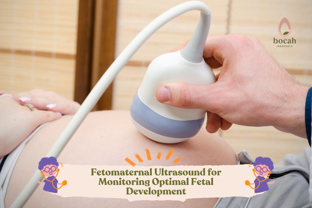
The fetomaternal ultrasound procedure is performed similar to transabdominal ultrasound, which involves applying gel on the abdomen, and then the doctor will move the ultrasound transducer around the abdominal area.
This examination is done to determine the blood circulation in the fetal body, uterus, and placenta. Additionally, this examination can be used to indicate whether the fetal organs receive an adequate blood supply.
Mulai Promil Sekarang
There are several indications when a mother may need this examination, such as:
Delayed fetal development during pregnancy being carried out
Reduced fetal movement
Pregnancy with twins
Twin to twin transfusion syndrome (TTTS) symptoms
A history of miscarriage
Risk of diabetes or preeclampsia conditions
Presence of rhesus factor abnormalities
Smoking habit before pregnancy
Types of Fetomaternal Ultrasound
Fetomaternal ultrasound examination also comes in several types, such as:
Uterine blood vessel Doppler ultrasound
This examination aims to ensure that the uterine arteries supplying blood to the uterus are sufficient. Typically, this examination is performed for mothers at risk of preeclampsia. This is because preeclampsia conditions will affect the performance of blood vessels that deliver oxygen and nutrients to the uterus.
Umbilical artery Doppler ultrasound
This examination aims to observe the blood flow from the placenta to the fetus through the umbilical cord. This is done to ensure that the fetus receives the necessary substances from the placenta. Typically, this ultrasound is recommended for pregnant mothers carrying twins.
Middle cerebral artery Doppler ultrasound
This middle cerebral artery Doppler ultrasound examination is used to observe blood flow in the fetal brain. Typically, this examination is performed if any issues are found during the umbilical artery Doppler examination.
Additionally, middle cerebral artery Doppler ultrasound is also used to determine the possibility of fetal anemia due to rhesus factor abnormalities. It also assesses the organ response in cases of slow-growing fetuses (IUGR).
Ductus venosus Doppler ultrasound
The ductus venosus Doppler ultrasound examination is performed when there is a fetus with slow development, and it assesses blood flow in the fetal heart. This examination is also conducted to ensure that there is no pressure on the fetal heart due to its slow growth.
When Can Fetomaternal Ultrasound Be Performed?
Fetomaternal ultrasound examination can be performed when the gestational age reaches 8-10 weeks. Although it uses sound wave technology, this ultrasound is safe and does not cause discomfort.
Basically, this examination is safe for pregnant mothers as long as it is adjusted to their individual conditions. Therefore, mothers do not need to worry if they need to undergo fetomaternal ultrasound examination.
Interested in articles related to fertility, pregnancy programs, and other pregnancy information? Stay tuned for more updates on Bocah Indonesia.
This article has been medically reviewed by Dr. Chitra Fatimah.
Source:
Feucht, U., et al. (2021). The ability of continuous-wave Doppler ultrasound to detect fetal growth restriction. PLoS One. 2021 Aug 9;16(8): e0255960. https://pubmed.ncbi.nlm.nih.gov/34370790/
Oglat, A.A., et al. (2018). A Review of Medical Doppler Ultrasonography of Blood Flow in General and Especially in Common Carotid Artery. J Med Ultrasound. 2018 Jan-Mar; 26(1): 3–13. https://www.ncbi.nlm.nih.gov/pmc/articles/PMC6029191/