Common Tubal and Uterine Abnormalities Found in HSG Examination
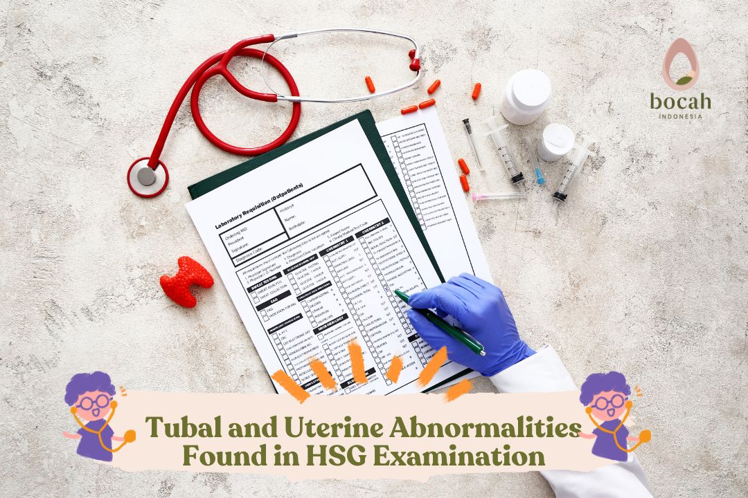
Hysterosalpingography (HSG) is an outpatient radiological procedure to evaluate the uterine cavity and the presence of blockages in the fallopian tubes. This examination is commonly performed as part of infertility evaluation.
Hysterosalpingography (HSG) is a radiological procedure performed to visualize the interior of the uterus and the patency of the fallopian tubes (whether there are blockages in the fallopian tubes). This procedure is usually a follow-up to evaluate the causes of infertility in women after pelvic ultrasonography (USG) examination. In the HSG procedure, a contrast agent is injected into the uterus and then visualized with X-rays.
In normal conditions, the fallopian tubes will appear as thin, smooth lines that widen at the ampullary part. This procedure is also beneficial for evaluating abnormalities inside the uterine cavity. Abnormal findings from HSG are followed up with hysteroscopy procedures to confirm and, if possible, address existing abnormalities.
The HSG procedure is usually performed between the 6th and 11th days from the first day of menstruation to avoid the possibility of pregnancy and menstrual bleeding.
Common Abnormalities Found in HSG Procedures
In HSG procedures, a thin tube is inserted through the vagina and cervix. Then, a contrast agent is injected into the uterus through that tube. Subsequently, a series of X-ray images or fluoroscopy will follow the movement of the contrast agent into the uterus and then into the fallopian tubes. This contrast agent will appear white on the screen.
Tanya Mincah tentang Promil?
If there are abnormalities in the uterine wall or the shape of the uterus, they will be depicted. If the fallopian tubes are open, then the contrast agent will gradually fill them. The contrast agent will then “spill” into the pelvic cavity, where the body absorbs it. The filling and draining pattern of this contrast agent will provide information about abnormalities in the uterus and fallopian tubes.
Below are some abnormalities in the fallopian tubes and uterus commonly found through the HSG procedure.
A. Abnormalities in the Fallopian Tubes
Tubal abnormalities observed in HSG can be congenital, due to spasms, occlusion, or infection.
- Tubal blockage. The presence of blockages in the tubes is confirmed by the absence or only partial filling of the fallopian tubes with contrast agent. The most common causes of tubal blockage are pelvic inflammatory disease, history of ectopic pregnancy, and endometriosis.
- Hydrosalpinx. This condition is defined as the dilation of the fallopian tubes due to fluid accumulation and is most often caused by peritubal adhesions. In cases of hydrosalpinx on HSG, the fallopian tubes are filled with contrast and dilated, but without any “spill” into the pelvic cavity.
- Peritubal adhesions. In a
- normal HSG, the contrast agent will freely spill from the fallopian tubes into the pelvic cavity. In cases of peritubal adhesions, the spillage appears as a contrast locus (irregular white area) near the ampullary end of the tube. This is the most common site of peritubal adhesions.
B. Uterine Abnormalities
Uterine abnormalities through the HSG procedure are generally divided into 2:
- Filling defects in the uterus
Possible causes of filling defects in the uterus by contrast agents include polyps, endometrial hyperplasia (thickening of the uterine wall), fibroids, uterine adhesions, and septate uterus.
- Endometrial polyps. The filling defect in this abnormality is clearly visible and most prominent in the early filling phase. Small polyps may not be visible if the contrast agent adequately distends the uterine cavity. Additionally, in HSG, polyps can be difficult to differentiate from fibroids, thus further examination through SIS (saline infusion sonohysterography) or hysteroscopy may be necessary.
- Uterine fibroids. Submucosal fibroids appear as filling defects on HSG, which can also enlarge the uterine cavity. As a result, small fibroids may not be visible. In rare cases, fibroids may occlude a portion of the fallopian tube, making contrast agent filling appear inadequate.
- Uterine adhesions. These adhesions are often caused by a history of injury to the uterine cavity, such as due to curettage or infection. Typically, HSG shows a distorted uterine contour with irregular filling defects. In women with Asherman’s syndrome (uterine adhesions accompanied by amenorrhea and infertility), the uterine cavity may be partially or completely obstructed by adhesions.
- Septate uterus. The presence of a septum or dividing wall in the uterus is an abnormality. This condition can lead to miscarriage but can be corrected through operative hysteroscopy.
- Uterine contour abnormalities
Uterine contour abnormalities include all types of abnormalities in the contour of the uterine wall or shape of the uterus. Some causes include:
- Adenomyosis. This is a condition where endometrial glands appear within the uterine muscle layer (myometrium). As a result, the uterine muscle layer enlarges and thickens. On HSG, adenomyosis appears as an enlarged cavity with clusters of contrast-filled sacs extending beyond the normal endometrial layer boundaries. Thus, the contrast agent appears to fill into the myometrial layer. Adenomyosis can appear diffuse (widespread) or focal (only in certain places).
- Congenital uterine abnormalities. Congenital uterine abnormalities arise from abnormalities in the Mullerian ducts, which are precursors to the female reproductive tract. HSG can identify various types of congenital uterine shape abnormalities, including unicornuate uterus (left), bicornuate uterus (middle), and arcuate uterus (right). However, to confirm the diagnosis of these abnormalities, better visualization of the outer uterine contour is needed through magnetic resonance imaging (MRI) examination.
- Exposure to diethylstilbestrol (DES). On HSG, the uterus exposed to synthetic estrogen DES appears as a “T-shaped configuration” in about one-third of cases. This T-shaped appearance is caused by the shortened upper segment of the uterus.
Diagnostic Accuracy of HSG Procedure
The accuracy of HSG examination results is influenced by many factors, such as underlying abnormalities, the skill and experience of the performing and interpreting physician, and the standards used to validate HSG results. Essentially, this procedure is most beneficial for assessing tubal blockages.
Studies have found that in cases of tubal blockages, compared to other examination modalities, HSG provides accuracy of up to 90 percent. In contrast, for uterine abnormalities, the accuracy of HSG examination is only about 70 percent, so findings need to be further confirmed using other examination modalities.
Conclusion
Hysterosalpingography (HSG) is a non-invasive and relatively low-cost diagnostic procedure compared to laparoscopy and other modalities. To date, HSG is still recognized as quite accurate in diagnosing abnormalities in the female reproductive tract. If HSG indicates tubal blockage or uterine abnormalities, the doctor may suggest additional procedures such as hysteroscopy (uterine endoscopy) or laparoscopy to confirm the diagnosis and address the issue.
This article has been medically reviewed by Dr. Chitra Fatimah.
Source:
- American College of Obstetricians and Gynecologists. [Last reviewed April 2023]. FAQ143. Hysterosalpingography. URL: https://www.acog.org/womens-health/faqs/hysterosalpingography#:~:text=Hysterosalpingography%20(HSG)%20is%20an%20X,a%20normal%20size%20and%20shape.
- American Society of Reproductive Medicine. Hysterosalpingogram (HSG). [Revised 2015]. URL: https://www.reproductivefacts.org/news-and-publications/patient-fact-sheets-and-booklets/documents/fact-sheets-and-info-booklets/hysterosalpingogram-hsg/.
- Banotra P, Ahmad Z. Diagnostic accuracy of hysterosalpingography and laparoscopy in tubal patency. International Journal of Reproduction, Contraception, Obstetrics and Gynecology. 2022 Feb 1;11(2):543-7.
- Lee SI, Kilcoyne A. Hysterosalpingography. In: UpToDate, Post, TW (Ed), UpToDate, Waltham, MA, 2022.
- Mayer C, Deedwania P. Hysterosalpingogram. [Updated 2022 Jan 24]. In: StatPearls [Internet]. Treasure Island (FL): StatPearls Publishing; 2022 Jan-. URL: https://www.ncbi.nlm.nih.gov/books/NBK572146/.
- Radiology Info. Hysterosalpingography. [Last reviewed 2019 Nov 25). URL: https://www.radiologyinfo.org/en/info/hysterosalp.


